
Blue Eyes and Light Sensitivity: True or False? Debunking Myths About Light Eyes
Are people with blue eyes really more sensitive to light? It’s one of the most persistent beliefs about eye color — and like many popular beliefs, it contains a kernel of truth wrapped in significant misunderstanding. At FLAAK, where we specialize in keratopigmentation and corneal science, we receive this question regularly from patients considering a permanent eye color change. Here is the complete, science-based answer — and what it means for anyone exploring eye color transformation in Paris.
The Short Answer: Partly True, Often Exaggerated
Blue eyes do, on average, experience greater light sensitivity than dark eyes — but the difference is more nuanced than most articles suggest, and it is certainly not a fixed biological destiny. Understanding why requires a brief look at iris anatomy and the role of melanin in the cornea and iris.
Melanin, Iris Color, and Light Filtering
The color of your iris is primarily determined by the concentration of melanin — the same pigment responsible for skin and hair color. Dark eyes (brown, black) contain high concentrations of melanin in the iris’s anterior stromal layer. Blue eyes contain very little melanin; the blue color we perceive is largely a structural phenomenon caused by light scattering (Tyndall effect) through a virtually depigmented iris stroma.
Melanin’s primary optical function in the eye is to absorb light. A densely pigmented iris acts as a biological light filter — absorbing stray photons that would otherwise scatter within the eye and create glare or wash out visual contrast. A lightly pigmented iris transmits more of this scattered light, which can manifest as increased sensitivity to bright conditions.
Myth 1: Blue Eyes Are Always More Light-Sensitive
Verdict: Partly true, but individual variation matters enormously.
The statistical correlation between low iris melanin and increased photophobia (light sensitivity) is real but modest. Studies measuring pupillary light reflex and subjective photophobia scores across eye colors do find that blue-eyed individuals report higher sensitivity on average — but the standard deviation within any eye color group is large. Many blue-eyed people report no unusual sensitivity to light whatsoever, while some brown-eyed individuals are highly photophobic due to neurological or corneal causes that have nothing to do with iris pigmentation.
Factors that influence light sensitivity far more than iris color include:
- Pupil size and reactivity (neurological factors)
- Corneal surface health (dry eyes, scarring)
- Migraine disorder (a major driver of photophobia)
- History of corneal or retinal procedures
- Medications affecting pupil dilation
- Ambient lighting adaptation habits
Myth 2: Changing Eye Color with Keratopigmentation Increases Light Sensitivity
Verdict: False, when performed correctly.
This is a concern FLAAK hears regularly from patients considering keratopigmentation. The answer lies in understanding exactly where the biocompatible pigments are deposited during the procedure.
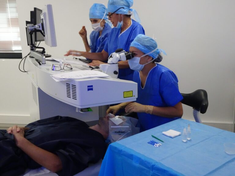
At FLAAK, the VisuMax femtosecond laser creates a precise intrastromal pocket in the peripheral cornea — outside the optical zone. The pigment does not enter the anterior chamber, does not contact the iris, and does not obstruct the pupil. It adds an optical layer of color in the stroma that mimics the visual appearance of a darker iris without altering the actual pupillary aperture or the light-filtering properties of the iris itself.
Because the pigment is placed peripherally, it does not reduce light transmission through the optical center of the cornea. FLAAK’s post-operative data shows no statistically significant change in photophobia scores between pre- and post-operative assessments when correct surgical technique is used.
Myth 3: Light-Eyed People Should Avoid Eye Surgery
Verdict: False.
Blue and green-eyed patients are among the most common candidates for keratopigmentation at FLAAK — precisely because they want to add iris depth and color richness that their natural pigmentation does not provide. The pre-operative assessment conducted by our ophthalmologist includes a full evaluation of corneal health, intraocular pressure, and endothelial cell count — none of which are meaningfully correlated with iris color.
A blue-eyed patient with a healthy cornea is an excellent candidate for keratopigmentation. A blue-eyed patient with thin corneas or elevated intraocular pressure is not — and will be told so clearly by our ophthalmologist, regardless of how much they may want the procedure.
Myth 4: Darker Eyes After Keratopigmentation Will Reduce Light Sensitivity
Verdict: Not significantly.
Some patients hope that changing their eye color to a darker shade through keratopigmentation will reduce their existing photophobia. This expectation should be managed carefully. Because the pigment is placed in the corneal stroma rather than in the iris itself, it does not replicate the full light-filtering effect of a naturally dark iris. The iris’s melanin absorbs intraocular scattered light — a function that corneal stroma pigment does not meaningfully replace.
Patients with clinical photophobia should address that condition through appropriate neurological or ophthalmological treatment, not through keratopigmentation. FLAAK is a cosmetic-surgical procedure for eye color change — it is not a treatment for photophobia.
Myth 5: Albinism and Blue Eyes Are the Same
Verdict: Completely false.
Albinism is a genetic condition characterized by the complete or near-complete absence of melanin throughout the body — including the retina, iris, and skin. People with albinism typically have very pale or pink-appearing eyes (due to visible blood vessels through unpigmented tissue) and severe photophobia caused in part by foveal hypoplasia and retinal underdevelopment.
This is categorically different from blue eyes, which have reduced but not absent iris melanin and no retinal development abnormality. Blue-eyed individuals do not have albinism, do not have the retinal anomalies associated with albinism, and their light sensitivity — to whatever degree it exists — operates through a completely different mechanism.
The Science of Iris Color Change and Corneal Optics
Understanding how the cornea and iris interact optically is important context for anyone considering keratopigmentation. Light enters the eye through the cornea, passes through the pupil, and strikes the retina. The iris — the ring of tissue surrounding the pupil — controls how much light enters by expanding or contracting the pupillary aperture.
The cornea’s primary optical function is refraction — bending incoming light to focus it on the retina. The corneal stroma itself is largely transparent; it does not significantly filter or absorb light under normal conditions. The biocompatible pigments introduced during keratopigmentation add a visible color layer in the peripheral stroma, creating the appearance of a different iris color when viewed from the outside — but this layer does not meaningfully alter the optical path of light toward the retina for the patient.
Practical Recommendations for Light-Sensitive Patients at FLAAK
For patients who do experience genuine light sensitivity and are also interested in keratopigmentation, FLAAK recommends the following approach:
- Rule out underlying causes first. See a neurologist or specialist to determine whether your photophobia has a treatable cause (migraine, dry eye disease, anterior uveitis) before considering any surgical eye color change.
- Bring all records to your FLAAK consultation. Our ophthalmologist will factor any documented photophobia history into the candidacy assessment.
- Set realistic expectations. Keratopigmentation will change how your eyes look — it will not change how much light your retina receives or processes.
- Post-operative adaptation period. All patients experience some light sensitivity in the first 7–14 days post-keratopigmentation as the cornea adapts. This is temporary and expected — not a complication.
Blue Eyes and Keratopigmentation: Who Comes to FLAAK?
A significant proportion of FLAAK’s patients are blue or green-eyed individuals from across Europe who wish to permanently change their eye color to darker shades — amber, brown, dark grey, or black. For these patients, keratopigmentation offers what no other method can: a true structural change to apparent iris color that is permanent, does not involve a foreign body inside the eye (unlike iris implants), and does not destroy iris tissue (unlike laser depigmentation).
The procedure is performed in Paris by FLAAK’s ophthalmologist, using the VisuMax femtosecond laser and certified biocompatible pigments. Price starts at €5,500 for both eyes, inclusive of pre-operative assessment and post-operative follow-up.
Key Takeaways
- Blue eyes are statistically somewhat more light-sensitive than dark eyes due to lower iris melanin — but individual variation is large and other factors dominate
- Keratopigmentation at FLAAK does not increase light sensitivity because the pigment is placed in the peripheral corneal stroma, outside the optical zone
- Keratopigmentation does not meaningfully reduce existing photophobia — it is a cosmetic procedure, not a treatment for light sensitivity disorders
- Blue-eyed patients are excellent candidates for keratopigmentation if their corneal health parameters are within safe ranges
- The VisuMax femtosecond laser ensures that the intrastromal pocket is created without disrupting the corneal optical zone
Safety and keratopigmentation protocol
Keratopigmentation is performed by a qualified ophthalmologist in a sterile medical environment in Paris. The procedure uses the VisuMax Zeiss femtosecond laser, a cutting-edge technology that creates an intracorneal tunnel with micrometric precision. The biocompatible pigments used are CE-certified and specifically designed for ophthalmic use, ensuring optimal tissue tolerance.
Unlike iris implants or permanent colored contact lenses, keratopigmentation does not affect the internal structures of the eye. The pigment remains confined within the corneal thickness, with no contact with the crystalline lens or the anterior chamber. This approach significantly minimizes risks of intraocular inflammation, glaucoma, or cataracts. The entire procedure takes approximately 30 minutes under topical anesthesia, completely painless.
A comprehensive preoperative assessment including corneal topography, pachymetry, and fundus examination is systematically performed before any intervention. This assessment confirms patient eligibility and allows personalization of the desired eye color change through a realistic 3D simulation.